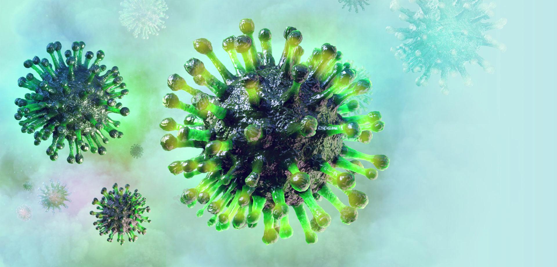
How does a COVID-19 infection develops: un update
Red blood cells transport oxygen from the lungs to all organs and the rest of the body. Red blood cells contain haemoglobin, a protein that consists of four groups of heme. These four groups have iron ions trapped in their centres, with the groups of heme acting as a container so the ions can be transported safely. Iron in its free form is quite toxic. Haemoglobin is used to bind to oxygen as it enters the lungs.
When red blood cells reach the lung alveoli, where gas exchange takes place, the iron ions switch between the 2+ and 3+ ionic states and bind to oxygen, then transport oxygen to where it is needed in the body.
COVID-19 binds to the hemoglobin beta chain
The hemoglobin molecule consists of four porphyrin chains, two of which are identical. The most common hemoglobin in humans consists of two alpha and two beta chains. The COVID-19 virus binds to the beta chains, causing the blood cells to produce glycoproteins. These glycoproteins bind to the haem, causing it to release the iron ion. Without its iron ion, oxygen can no longer bind to the haemoglobin molecule and therefore it is released. In the long term, this causes oxygen saturation to decease: the SpO2 level drops.
The release of erythropoietin in the kidneys
The body compensates for this lack of ability to transport and release oxygen (hypoxia) by allowing the kidneys to release hormones such as erythropoietin. Erythropoietin increases the production of new red blood cells with haemoglobinis released in the bone marrow. Therefore, increased haemoglobin, reduced oxygen saturation in the blood and increased iron are some of the most important characteristics in Covid-19 infection. Free iron from the infected blood cells entering the blood is highly reactive and causes oxidative damage. There are two ways in which the lungs can eliminate free iron. Firstly, the alveoli contain macrophages that can detect and absorb free radicals such as free iron. Secondly, epithelial surface linings contain a thin layer of liquid rich in antioxidant molecules, which are able to bind free iron.
Pulmonary oxidative stress
Due to the excess iron, these mechanisms soon become overloaded and pulmonary oxidative stress begins. This leads to damage and inflammation, which can be seen in CT scans of the lungs of COVID-19 patients, in which the fluid layer drops into the alveoli. In pneumonia infections, this damage is almost always in only one lung, but in COVID-19 patients, it is in both.
The red blood cells are no longer able to transport oxygen. The affected haemoglobin molecules are permanently deprived of their ability to transport oxygen, since they have lost their iron ions.
The liver is trying to store iron
The liver attempts to remove the excess iron from the blood and to store it, but the liver also becomes overloaded due to the lack of oxygen. Liver cells die, causing alanine aminotransferase (ALT) and other liver enzymes to be released. Increased ALAT, CRP and Alkalic Phosphatase are also characteristic.
Release of cytokines
The cytokine Interleukin-8 is released in the upper respiratory tract in response to a viral respiratory infection, and polymorphonuclear neutrophils play an important role in this inflammatory reaction by recreation of ENS. This is in contrast to the predominantly eosinophilic reaction that occurs in atopic diseases (allergies) of the upper and lower respiratory tract.
Effect on HPU (under construction)
If you, as a patient with HPU, have questions, you can send an e-mail to info@nullkeac.nl.